some radiology for pediatricians
Модераторы: Ren_Yumi, Alon, dr.Ira
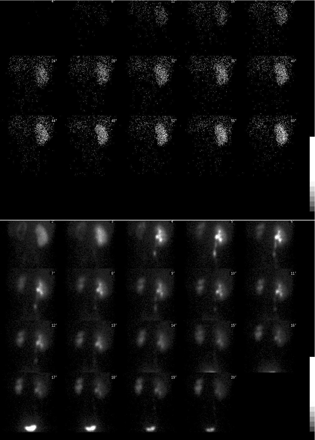
Меккелев дивертикул?Ren_Yumi писал(а):Четырехлетний мальчик с кровотечением per rectum:
Но не потому, что я поняла на что смотрю (про то, что вижу я и не говорю), а потому, что латинские буквы знаю
Так что можно сначала для нас, участковых, рассказать - что, где и как исследуется.
Алла
"Life is either a daring adventure, or nothing."
"Life is either a daring adventure, or nothing."
Meckel's Diverticulum
Clinical:
A Meckel's diverticulum represents a persistence of the omphalomesenteric (vitelline) duct at its junction with the ileum (the diverticulum therefore arises from the anti-mesenteric border). Meckel's are found in about 2% of the population and the great majority of patients are asymptomatic (80%). Ectopic mucosa is found in about 60% of Meckels and it is most commonly gastric (about 90% of cases) [1]. If symptomatic, patients usually present by age 2 years (60% of cases) and symptomatic lesions are more common in males (3:1). Symptoms in children include painless blood per rectum (most common manifestation), while adults most often present with Meckel's diverticulitis. Meckel's are typically located approximately 2 feet from the ileocecal valve.
Complications are seen in 20% of patients and include:
Bleeding (most common complication): Generally more common in those lesions lined by gastric mucosa (found in 95% of bleeding lesions). Ectopic gastric mucosa is found in 15-25% of Meckel's, but in about 50% of symptomatic patients [2].
Intussusception, Volvulus
Diverticulitis
Radiographic and Nuclear Medicine Exam Findings:
Radiographic findings on the barium exam include a wide mouth diverticulum which contains a normal small bowel fold pattern.
Tc99m-pertechnetate is the agent used for scintigraphic imaging as pertechnetate will accumulate within the mucous producing cells of the gastric mucosa. A dose of 10mCi (370 Mbq) IV (200uCi/kg in children) is used. An area of at least 2 cm2 of mucosa is necessary for successful scintigraphic visualization. Sensitivity for the exam is over 80-90% in children, about 60% in adults (symptoms in the adult age group are usually unrelated to the presence of ectopic gastric mucosa). Gastric concentration usually begins 5 to 10 minutes post injection, and then increases in intensity over time. Ectopic gastric mucosa demonstrates a similar pattern of tracer accumulation.
A positive scan demonstrates a focal area of increased activity in the RLQ which appears at the same time as the stomach and increases in intensity in a similar manner. On the lateral view, the activity should be unrelated to ureteral activity.